
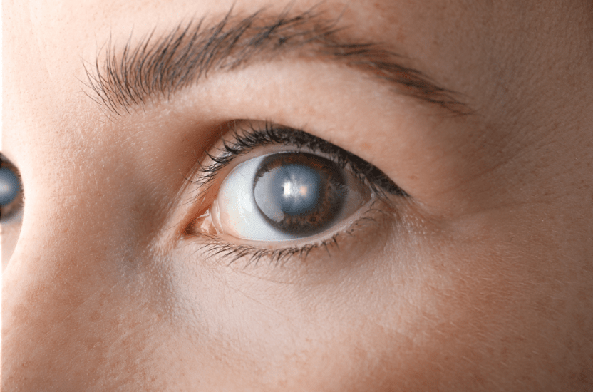
What is glaucoma?
Glaucoma is a chronic, progressive eye disease in which the optic nerve is gradually damaged, usually due to increased intraocular pressure (TOT). If glaucoma is not treated, it can lead to irreversible vision loss - up to complete blindness.
- Safe and comfortable treatment
- Caring for children and adults
- Service and availability
- International standards
- Individual approach
Answers to questions
• Fluid (moisture) is constantly produced inside the eye.
• If it is not removed properly, the pressure in the eye increases.
• Increased pressure compresses the optic nerve, disrupts its nutrition and function.
• There are defects in the field of vision that do not return, even if the pressure is reduced.
Type of glaucoma and features:
Open form: The most frequent. It proceeds gradually, without pain.
Closed-angle: Can develop suddenly, with severe pain and rapid loss of vision.
Congenital glaucoma: Found in infants or children.
Normotensive: Damage to the optic nerve at normal pressure.
Symptoms of glaucoma:
In the initial stages (especially the open form):
- No symptoms or only mild vision loss
- Gradual loss of lateral (peripheral) vision
In acute attack (closed-angle glaucoma):
- Sharp pain in the eye and head
- Redness of the eye
- Fog in the eyes, rainbow circles around the light
- Nausea, vomiting
How glaucoma is diagnosed:
- Measurement of VOT (tonometry)
- Optic nerve examination (ophthalmoscopy)
- Checking the field of view (perimetry)
- OCT (optical coherence tomography) - detailed assessment of the optic nerve
Treatment:
- Pressure-reducing eye drops (most common)
- Laser treatment (e.g. laser trabeculoplasty)
- Microsurgical surgery (if other methods do not help)
SUMMARIZE:
Glaucoma is a silent "killer of vision," because it often proceeds painlessly and imperceptibly.
Vision lost due to glaucoma cannot be restored.
Regular preventive examinations - especially after 40 years - give a chance to detect the disease in time.
People of any age can get glaucoma, but most often - after 40 years. The risk increases with age, especially after 60-70 years.
The main age categories:
Congenital glaucoma
- A rare but very serious form.
- It is detected in newborns or in the first years of life.
- Causes - congenital disorders of the outflow of intraocular fluid.
Juvenile glaucoma
- Develops in children and adolescents under 35 years of age.
- Often has a genetic nature.
Young glaucoma
It may appear after 30 years, especially with hereditary predisposition, myopia, endocrine disorders, eye injuries, etc.
The most common is primary open-label glaucoma
- It begins after 40 years (in 1-2% of people).
- After 60 years - the risk increases to 4-10%.
- At the age of 80 + - up to 15-20% of people have signs of glaucoma.
Age-related risk factors:
- Age > 40 - the main factor
- Heredity (glaucoma in parents/grandparents)
- Diabetes mellitus, hypertension, atherosclerosis
- Nearsightedness or farsightedness
- Long-term use of corticosteroids
Recommendations:
After 40 years - 1 preventive examination by an ophthalmologist every year.
If there is heredity or other risks - an examination every 6-12 months, even without complaints.
People suffer from glaucoma mainly due to a violation of the outflow of intraocular fluid, which leads to an increase in intraocular pressure (TOT) and damage to the optic nerve. This is a chronic disease that develops gradually and often imperceptibly.
The main causes of glaucoma:
1. Impaired circulation of intraocular fluid
- Normally, the fluid is constantly produced and flows out of the eye.
- If the drainage system (trabecular network) is blocked or narrowed, the fluid accumulates - the pressure rises - the optic nerve is squeezed.
2. Heredity
- If one of the relatives has glaucoma, the risk increases 4-10 times.
- This is one of the most important reasons why you should regularly check your eyesight after 40 years.
3. Age changes
After 40-45 years:
- reduced efficiency of liquid outflow;
- eye tissues lose elasticity;
- the risk of UTI increase increases.
4. Ocular comorbidities
Myopia, cataracts, eye inflammation (uveitis), eye injuries.
5. Long-term corticosteroid use
Eye drops, inhalers, ointments - can cause steroid glaucoma.
6. Common diseases of the body
- Hypertension
- Atherosclerosis
- Diabetes mellitus
- Migraine
- Cerebrovascular accident
7. Stress and circulatory disorders
Poor blood supply to the optic nerve makes it more vulnerable to increased pressure.
Additional risk factors:
Factor/Risk
Age 60 +: Significantly increasing
Dark iris color: Higher frequency of glaucoma
Asian or African origin: More likely
Smoking: Weakens the blood supply to the optic nerve
Sitting in the dark for a long time: Can cause an attack of closed-angle glaucoma
SUMMARIZE:
Glaucoma occurs due to impaired drainage
intraocular fluid and hereditary or age predisposition.
The main danger is a slow, asymptomatic loss of vision.
Regular check-ups after age 40 are key to
early detection.
It is possible to detect glaucoma at an early stage, when it does not yet affect vision, only during a preventive examination by an ophthalmologist. In most cases, glaucoma is asymptomatic, so regular diagnosis is the only way to detect it in time and preserve vision.
How exactly glaucoma is detected at the preclinical stage:
1. Measurement of intraocular pressure (tonometry)
- The main method of primary detection.
- Normal pressure: 10-21 mm Hg Art.
- Increased pressure is one of the first signs of glaucoma, although not always (there is glaucoma with normal pressure!).
2. Optic nerve examination (ophthalmoscopy)
- The doctor assesses the shape, color and degree of "excavation" (excavation) of the optic disc.
- Initial changes are visible even before symptoms appear.
3. Checking visual fields (perimetry)
- Reveals invisible to the patient "fallout" in the side star, which are not noticed in everyday life.
- This is a sign of the initial damage to the optic nerve.
4. OCT (optical coherence tomography)
- The most accurate method of early diagnosis.
- Allows you to measure the thickness of the nerve fibers of the retina - with glaucoma, they gradually become thinner.
5. Gonioscopy
Examination of the angle of the anterior chamber of the eye - allows you to identify the risk of closed-angle glaucoma even before the attack.
Who and when to be examined?
Risk group/Recommended frequency of examinations
Persons after 40 years: 1 time in 1-2 years
People with a family history of glaucoma: Every year
Patients with diabetes mellitus, hypertension: Every year
Individuals with myopia/farsightedness: Every year
SUMMARIZE:
Glaucoma can begin to destroy the optic nerve before a person notices vision problems.
It can be detected at an early stage only through an ophthalmic examination.
Timely diagnosis is the only chance to preserve vision for life.
To check whether there is glaucoma, it is necessary to undergo a special ophthalmological examination. Glaucoma can develop without obvious symptoms, so it is impossible to independently detect it in the early stages.
The main methods for checking glaucoma:
1. Tonometry - measurement of intraocular pressure (TOC)
- One of the first and most important tests.
- Normal values: 10-21 mm Hg Art.
- But: glaucoma can be at normal pressure (normotensive glaucoma!).
2. Ophthalmoscopy (optic disc examination)
The doctor checks whether there are changes in the structure of the optic nerve - "excavation" (excavation), which indicates glaucoma.
3. Perimetry (visual field examination)
It detects invisible "dips" in the side star that occur when the optic nerve is damaged.
4. OCT (optical coherence tomography)
- The most accurate method of assessing the thickness of the retinal fibers and the state of the optic nerve.
- Allows you to detect glaucoma changes even before the onset of symptoms.
5. Gonioscopy
Examination of the anatomy of the angle of the anterior chamber of the eye is important for the detection of closed-angle glaucoma.
Comprehensive diagnosis of glaucoma usually includes:
- Collection of history (is there glaucoma in the family?)
- Measurement of VOT (tonometry)
- Optic disc examination (ophthalmoscopy)
- Checking the field of view (perimetry)
- OCT (as indicated)
- Corneal thickness measurement (pachymetry) - for
When to check?
Patient category/Frequency of examination
All people after 40 years: Once every 1-2 years
At risk (heredity, diabetes, migraine, hypertension, steroids): Every year or more often
SUMMARIZE:
It is possible to check whether there is glaucoma only with an ophthalmologist using special diagnostic methods.
The sooner it is detected, the more likely it is to preserve vision.
more accurate pressure estimation
Manifestations of glaucoma depend on its type and stage. The most dangerous thing is that in the early stages of glaucoma can proceed without symptoms, and the patient learns about the disease only when there is already damage to the optic nerve.
The main manifestations of glaucoma:
In open-angle glaucoma (the most common form):
- There is no pain or redness, and that is why she is called the "quiet thief of vision."
- Gradual, imperceptible loss of peripheral (lateral) vision.
- Vision as if "narrows into a tunnel."
- Deterioration of twilight vision.
- A feeling of slight blurred vision.
- Often - there are no complaints at all until the late stages.
In closed angle glaucoma (acute attack):
- Sharp, severe pain in the eye, which can give to the head.
- Redness of the eye.
- Blurred vision, "rainbow circles" around light sources.
- Nausea, vomiting.
- Significant reduction in vision for several hours.
This is an emergency - needs urgent intervention!
Common signs that can alert:
Symptom/What can mean
Gradual loss of lateral vision: Initial glaucoma
Eye pain, headache: Acute angle-closure glaucoma
Rainbow halos around the light: Increased pressure in the eye
Pressure, eye discomfort: Initial hypertension or glaucoma
Frequent visual impairment at dusk: Optic nerve disorder
Features of glaucoma:
- Vision damage is irreversible - lost vision is not restored.
- The only way to preserve vision is timely diagnosis and pressure control.
When to contact an ophthalmologist:
- After 40 years - annually.
- If there are relatives with glaucoma, diabetes, high blood pressure, eye injuries - review every year regardless of age.
The most effective methods of treating glaucoma are those that reduce intraocular pressure (IOP) and slow or stop damage to the optic nerve. It is impossible to completely cure glaucoma, but timely treatment helps to preserve vision for life.
The main methods of treating glaucoma:
Eye drops (conservative treatment)
This is the first line of therapy, especially in open-angle glaucoma.
Type of drops/Action
Beta-blockers: Reduce fluid formation
Prostaglandins: Improve fluid outflow
Carbonic anhydrase inhibitors: Reduce fluid production
Alpha-Adrenoreceptor Agonists: Dual Action: Production and Efflux
Combined drops: A combination of two mechanisms of action
IMPORTANT:
Drops are used constantly, even if there are no symptoms.
Violation of the regime is a threat of vision loss.
Laser treatment of glaucoma
Trabeculoplasty (SLT or ALT) - in open-angle glaucoma
- Improves the outflow of intraocular fluid.
- Outpatient, painless procedure.
Iridotomy (laser creation of an opening in the iris) - with closed-angle glaucoma
- Opens the angle for the outflow of liquid.
- Performed to prevent or treat acute attacks.
Surgical treatment (if drops and laser do not help)
Trabeculectomy
- Creating a new path for the outflow of fluid from the eye.
- One of the most effective methods for sustainable reduction of VOT.
Implantation of drainage devices
- For example, shunts (Ahmed, Baerveldt).
- Used for complex or secondary glaucomas.
Minimally Invasive Surgery (MIGS)
- The latest technology with less risk of complications.
- Often combined with cataract surgery.
Additional measures:
- Control of blood pressure, sugar, cholesterol.
- Avoiding stress and strong physical stress.
- Regular visit to the ophthalmologist (1-2 times a year or more often).
SUMMARIZE:
The most effective methods of treating glaucoma are those that provide control of TOT at the individual level.
The choice of method depends on the type of glaucoma, the degree of damage, age, general health.
Early detection + regular treatment = chance to save vision!
No, glaucoma cannot be completely cured because it is a chronic, progressive disease of the optic nerve. But it is possible to slow down its development and preserve vision if it is detected in time and properly treated.
What does it mean to treat glaucoma?
Vision lost due to glaucoma is not restored, since damage to the optic nerve is irreversible.
Treatment allows you to control intraocular pressure - the main cause of nerve damage.
With regular treatment, you can slow the progression of the disease.
Treatment Objective/Outcome
Lower BOT: Slow the progression of glaucoma
Stabilize the condition: Prevent loss of residual vision
Constantly monitor: Timely respond to changes
Why not delay:
- In glaucoma, changes in vision occur imperceptibly.
- If left untreated, the disease leads to total blindness.
- Regular use of eye drops, laser or surgical treatment is a vital support, not a temporary solution.
SUMMARIZE:
Glaucoma is not cured, but controlled.
With proper treatment, a person can maintain vision for life.
The main thing is timely diagnosis, discipline in treatment and regular examinations.