
What is diabetic retinopathy?
Diabetic retinopathy is a chronic complication of diabetes mellitus that affects the retina (retina) - the light-sensitive tissue lining the back of the eye. It occurs due to damage to the small blood vessels of the retina due to a prolonged increase in blood glucose levels.
Reasons:
- Prolonged course of diabetes mellitus (type 1 or 2)
- Uncontrolled blood sugar
- High blood pressure
- Kidney disease
- Smoking
- Safe and comfortable treatment
- Caring for children and adults
- Service and availability
- International standards
- Individual approach
Answers to questions
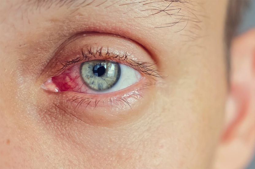
High glucose levels damage the retinal vascular walls, leading to:
• microaneurysms (protrusions of blood vessels),
• hemorrhages,
• leakage of fluid and fats,
• growth of new pathological vessels (neovascularization), which can cause retinal detachment or glaucoma.
Forms of diabetic retinopathy
1. Non-proliferative retinopathy (NPDR)
- early stage: microaneurysms, edema, hemorrhage
2. Proliferative retinopathy (PDR)
- severe stage: formation of new vessels, risk of blindness
3. Diabetic macular edema (DME)
- edema of the central retina (macula), the main cause of vision loss in diabetes
Symptoms
• In the early stages - absent
• Deterioration of vision, "veil," spots
• Reduced visual acuity
• Distortion of objects
• Sudden vision loss (with hemorrhage)
Diagnosis
• Examination of the fundus by ophthalmoscope
• Retinal photo (retinophoto)
• Fluorescein angiography
• OCT (optical coherence tomography)
Treatment
• Control of glucose, pressure, lipids
• Laser retinal coagulation
• Anti-VEGF injections (e.g. Aflibercept, Faricimab)
• Vitrectomy (surgery for complications)
Complications
• Retinal detachment
• Hemophthalmos (vitreous hemorrhage)
• Secondary glaucoma
• Blindness
Prevention
• Regular examinations by an ophthalmologist (every year, and with complications - more often)
• Effective control of glucose, blood pressure and cholesterol
• Quitting smoking
SUMMARIZE:
Diabetic retinopathy is a serious but controlled complication of diabetes.
Timely diagnosis is your best protection for preserving your vision.
Diabetic retinopathy is affected not so much at a certain age, but because of the duration of diabetes and the level of glucose control. However, there are general age trends:
General observations regarding age:
Type of diabetes/Age of onset of retinopathy/Comment
Type 1 diabetes: Usually 5-10 years after diagnosis, often after 20 years (in children rarely detected earlier)
Type 2 diabetes mellitus: Can be detected already at diagnosis (often in persons 40 + years old) (Often type 2 diabetes proceeds "quietly" until complications are detected)
Most often sick:
• Persons aged 40 to 70 years who have type 2 diabetes.
• Patients with type 1 diabetes mellitus who have been ill for more than 10-15 years.
Factors affecting the age of retinopathy:
• Duration of diabetes (key factor)
• Poor sugar control
• Hypertension
• Kidney disease
• Pregnancy (in women with diabetes can cause deterioration of the retina)
SUMMARIZE:
Diabetic retinopathy can occur at any age, but the risk increases significantly after 40 years or 5-10 years after the onset of diabetes, especially with poor control. That is why it is important to undergo regular retinal examinations even in the absence of symptoms.
Suffer from diabetic retinopathy due to damage to the blood vessels of the retina, resulting from prolonged elevated blood glucose levels. This is a complication of diabetes mellitus - both type 1 and 2.
The main causes and mechanisms of development:
1. Chronic hyperglycemia
High blood sugar levels damage the inner wall of the small capillaries (endothelium) that feed the retina. This results in:
- thinning and rupture of vessels,
- microaneurysms,
- retinal hemorrhages.
2. Circulatory disorders in the retina
- hypoxia (lack of oxygen) - the body stimulates the growth of new vessels (neovascularization). Newly formed vessels:
- brittle and brittle,
- cause bleeding and scarring,
- can exfoliate the retina.
3. Macular edema
Due to vascular damage, blood plasma seeps into the retinal tissues - edema of the central zone of vision (macula) - a decrease in visual acuity.
Risk factors contributing to diabetic retinopathy:
Factor/How it affects
Poor glucose control: The higher the HbA1c level, the greater the risk of vascular damage
Duration of diabetes: More than 5-10 years - significantly increases the likelihood of retinopathy
Hypertension: Increases pressure in the vessels of the eye, accelerates complications
Impaired lipid metabolism: Elevated cholesterol and triglycerides damage blood vessels
Smoking: Impairs blood circulation, promotes oxidative stress
Pregnancy: May temporarily increase retinopathy in diabetes
Genetic predisposition: Some patients have a higher sensitivity to diabetes complications
SUMMARIZE:
Diabetic retinopathy is affected due to a long-term metabolic disorder in diabetes that causes damage to the retinal vessels. The main "culprits" are high glucose levels, poor diabetes control, high blood pressure and time. But with proper control, you can significantly slow down or completely prevent the development of retinopathy.
Diabetic retinopathy in the early stages may not produce any symptoms and the patient may not yet experience blurred vision. That is why the only way to detect it at an early (asymptomatic) stage is a regular examination of the fundus by an ophthalmologist.
How to detect diabetic retinopathy before the appearance of complaints of vision?
1. Fundus examination (ophthalmoscopy)
• Performed after instillation of eye drops that dilate the pupil.
• The doctor can see:
o microaneurysms (first sign),
o minor hemorrhages,
o seal,
o leakage of liquid from vessels,
o Signs of macular edema.
2. Retinal photography (retinophoto)
• It is made by a special device - a foundation camera.
• Allows you to save images for later comparison and dynamic observation.
3. OCT (optical coherence tomography)
• A modern method to detect macular edema or structural changes in the retina even before vision deterioration.
• Botherless, fast research.
4. Fluorescein angiography
• Intravenous dye (fluorescein) is injected, retinal images are taken.
• Allows you to see the leaks, blocks of blood flow, areas of neovascularization.
• Used when proliferative stage is suspected.
Who and how often to undergo an examination of the fundus?
Patient Category/Frequency
Type 1 diabetes (5 years after diagnosis): Every year
Type 2 diabetes (since diagnosis): Immediately and annually
Pregnant women with diabetes Before pregnancy/in early stages and in each trimester:
In the presence of retinopathy Every 3-6 months (depending on the stage)
SUMMARIZE:
Diabetic retinopathy can progress without symptoms, so regular ophthalmological examination is the only way to detect the disease before vision loss. Early diagnosis allows you to start treatment in time and avoid blindness.
To check whether there is diabetic retinopathy, you need to contact an ophthalmologist who will conduct a specialized examination of the fundus. The following are key diagnostic methods that help detect retinopathy at any stage - even without symptoms.
How to check for diabetic retinopathy:
1. Fundus examination (ophthalmoscopy)
Mandatory procedure. After instilling drops that dilate the pupil, the doctor examines the retina. Detected:
- microaneurysms
- hemorrhages
- liquid leakage
- swelling
- pathological vessels (signs of proliferation).
2. Photo of the fundus (fundus photo)
- Gives a clear image of the retina.
- Stored to compare disease dynamics.
- You can spend in the office of an ophthalmologist or endocrinologist, if there is appropriate equipment.
3. OCT (optical coherence tomography)
• Modern technology of visualization of the retina in the section.
• Helps to identify:
o macular edema (even minor),
o change in retinal thickness,
o structural damage.
4. Fluorescein angiography
- Intravenous administration of contrast (fluorescein).
- Shows in detail the condition of the blood vessels:
blockages, fluid leaks, newly formed vessels.
- Applicable in complex or running cases.
5. Visual acuity measurement and macular function test
- Assistive techniques that can detect decreased vision even before imaging changes.
When to be examined:
Category of patients/When and how often to be examined
Type 2 diabetes: Immediately after diagnosis, then every year
Type 1 diabetes mellitus: 5 years after diagnosis, then every year
Pregnant women with diabetes: Before pregnancy or at its beginning, then - every trimester
Patients with detectable retinopathy: Every 3-6 months, depending on the condition
SUMMARIZE:
To check if there is diabetic retinopathy, be sure to consult an ophthalmologist who will examine the fundus, OCT, fundus photo or other tests. Early diagnosis avoids complications, including vision
Manifestations of diabetic retinopathy depend on the stage of the disease. In the early stages, it can be asymptomatic, so the patient does not notice deterioration of vision. However, with the progression of the disease, various visual symptoms appear.
The main manifestations of diabetic retinopathy:
In the early stages (non-proliferative retinopathy):
- There are no symptoms - often detected only during a preventive examination.
- Possible mild:
o blurred vision,
o sensation of tension in the eyes during loading.
In the middle and later stages:
- Blurred vision (veil on the eyes)
- Decreased visual acuity
- Spots, "flies," dark spots in sight
- Curvature of straight lines (sign of macular edema)
- Difficulty reading or recognizing faces
- Sudden loss of vision in one eye (may indicate vitreous hemorrhage or retinal detachment)
In diabetic macular edema:
- Decreased central vision
- Inability to clearly see objects directly in front of you
- Image distortion, "waviness" of lines
In proliferative retinopathy:
- Vision may deteriorate suddenly
- Frequent eye haemorrhages looking like dark spots or fog
- Possible complete blocking of vision in major hemorrhage
It is important to remember:
- The presence of diabetes even without vision problems is an occasion to regularly check the eye bottom.
- Early detection of retinopathy is a chance to preserve vision.
SUMMARIZE:
Manifestations of diabetic retinopathy range from complete absence of symptoms in the early stages to significant or even sudden deterioration of vision in the later stages. Therefore, regular preventive examinations by an ophthalmologist are critically important for preserving vision.
The most effective treatments for diabetic retinopathy depend on the stage of the disease and the presence of complications (for example, macular edema, vascular proliferation or hemorrhage). The main goal of treatment is to stop the progression of the disease, to preserve and improve vision.
1. Medical treatment (injections into the eye)
Anti-VEGF drugs are the most modern and effective method
These drugs block the vascular growth factor (VEGF), which stimulates the appearance of pathological vessels.
- Preparations:
Aylia (Aflibercept)
Wabismo (Faricimab)
- Indication: macular edema, proliferative retinopathy
- Effect: Reduced swelling, improved or stabilized vision
Corticosteroids (implants or injections)
• Reduce inflammation and swelling
• Used less frequently due to side effects (increased intraocular pressure, cataract development)
2. Laser treatment (photocoagulation)
Focal laser coagulation
- With macular edema
- Closes leaking vessels, stops fluid leakage
Panretinal laser coagulation
- In proliferative retinopathy
- Cauterizes the peripheral retina, reduces the need for oxygen - stops the growth of new vessels
- Objective: To prevent hemorrhage and retinal detachment
3. Surgical treatment (vitrectomy)
It is carried out in cases of:
- massive hemorrhages into the vitreous body,
- retinal detachment,
- fibro-vascular proliferation
During the operation:
- the vitreous body with blood is removed,
- the anatomy of the eye is restored,
- often combined with laser coagulation
Concomitant medication and prophylaxis:
- Blood sugar control (glycated hemoglobin HbA1c < 7%)
- Normalization of blood pressure and lipids
- Quitting smoking
- Regular ophthalmic examinations
SUMMARIZE:
The most effective treatments are anti-VEGF injections and laser coagulation.
In the later stages - vitrectomy.
But the key to successful treatment is diabetes control and regular follow-up, because early intervention avoids blindness.
Why can't retinopathy be "cured" completely?
- This is a chronic complication of diabetes.
- It occurs due to the constant influence of high glucose levels on the vessels of the retina.
- Even after successful treatment, the risk of repeated deterioration of vision remains if diabetes is not controlled.
What you can do:
In the early stages (non-proliferative retinopathy):
- Diabetes control (HbA1c < 7%)
- Normalization of pressure, cholesterol
- Healthy lifestyle
- Regular retinal examination - allows you to keep the disease under control without interventions
At the stage of complications:
- Anti-VEGF injections - can stop neovascularization and reduce swelling
- Laser treatment - prevents further damage to the retina
- Vitrectomy - with hemorrhage or detachment of the retina
IMPORTANT: These methods do not restore already dead retinal cells, but can stabilize or partially improve vision.
SUMMARIZE:
Diabetic retinopathy is incurable but very well controllable.
With timely detection and active treatment, you can completely stop the loss of vision and live a full life.
The key is regular checkups, glucose control and treatment in the early stages.