
What is age-related macular degeneration or AMD?
Age-related macular degeneration (AMD) is a chronic, progressive disease of the central part of the retina (the macula) that most often develops in people after the age of 50 and is one of the leading causes of central vision loss in old age. What is the macula? The macula is a small area in the center of the retina that is responsible for sharp central vision, necessary for reading, recognizing faces, driving, and more.
- Safe and comfortable treatment
- Caring for children and adults
- Service and availability
- International standards
- Individual approach
Answers to questions
Types of AMD:
Type/Description/Frequency
Dry (atrophic) form: Gradual degeneration of macular cells without the formation of new vessels. Vision deteriorates slowly. ~ 85-90% of cases
Wet (neovascular) form: Formation of pathological vessels under the retina that bleed and cause swelling. Vision is lost quickly. ~ 10-15% of cases, but causes ~ 90% of vision loss in AMD
Symptoms of AMD:
- Deterioration of central vision
- Difficult to read or recognize faces
- Curvature of straight lines (lines appear wavy)
- Darkened spot in the center of vision
- Color distortion
- Lateral vision is usually preserved
Risk factors:
- Age 50 + years
- Heredity (the presence of AMD in relatives)
- Smoking
- High blood pressure
- Poor nutrition (lack of antioxidants, omega-3)
- Obesity
- Excessive UV exposure (without glasses)
Diagnosis:
- Fundus examination
- OCT (optical coherence tomography) - visualizes the structure of the macula
- Fluorescein angiography - detects pathological vessels (in wet form)
- Amsler grid - test for curvature of straight lines (can be done at home)
Treatment:
Dry form:
- There is no specific treatment.
- Recommended:
o Intake of antioxidants (vitamins A, C, E, zinc, lutein, zeaxanthin)
o Balanced nutrition
o Control of pressure, weight, cholesterol
Wet form:
- Anti-VEGF injections into the vitreous:
o Aylia (Aflibercept)
o Lucentis (Ranibizumab)
o Bevacizumab
- Laser therapy (less commonly used)
- Photodynamic therapy
SUMMARIZE:
AMD is a serious age-related retinal disease affecting central vision.
Early diagnosis and regular observation can slow down the course of the disease, especially in the dry stage.
Injections of modern drugs with a wet form can preserve vision or even partially improve it.
Age-related macular degeneration (AMD) is a disease that is inherently associated with the aging of the body, so it most often occurs after 50 years.
At what age is AMD usually detected:
Age/Probability of AMD
Up to 50 years: Very rare, almost never happens
50-60 years: Low risk, but initial changes may already appear
60-75 years: Medium risk, frequency increases (especially dry form)
75 + years: High risk, highest number of cases, in particular wet form
Why does the risk increase with age?
- Reduced retinal blood supply
- Accumulated waste cell metabolism (druse) under the macula
- Photoreceptor cell repair is impaired
- Degraded antioxidant protection
Additional risk factors in old age:
- Smoking
- High blood pressure or cholesterol
- Unbalanced power supply
- Weak protective effect of retinal pigment layer
SUMMARIZE:
AMD is a disease of older people, most often occurring after 60 years of age, and the largest number of cases is in people over 75 years of age. Regular preventive examinations after 50 years help to identify the disease at an early stage.
Age-related macular degeneration (AMD) is affected due to a combination of age-related, genetic and external factors that lead to macular degeneration - the central part of the retina responsible for clear central vision.
The main reasons for the development of AMD:
🔹 1. Aging (age 50 +)
- The biggest factor.
- Over the years, the metabolism in the retina worsens, drusy (deposits) accumulate, which damage the macular cells.
🔹 2. Genetic predisposition
- If someone from close relatives had AMD, the risk of you increases 2-4 times.
- Mutations in genes (e.g. CFH, ARMS2) associated with inflammatory processes and oxidation in the retina have been identified.
🔹 3. Smoking
- One of the strongest external factors.
- Increases the risk of AMD in 2-4 times.
- Disrupts the blood supply to the retina and causes oxidative stress.
🔹 4. Excessive ultraviolet radiation
- Ultraviolet rays can damage retinal cells.
- Lack of sunglasses is an added risk.
🔹 5. Poor nutrition
- Lack of antioxidants (vitamins C, E, zinc, lutein, omega-3) weakens the protection of retinal cells.
- Excessive consumption of saturated fats - contributes to the accumulation of deposits.
🔹 6. Vascular disorders
High blood pressure, atherosclerosis, diabetes - impair blood circulation in the retina and accelerate macular degeneration.
🔹 7. Obesity, metabolic syndrome
Disorders of lipid metabolism contribute to the development of AMD and accelerate its transition into a wet form.
Risk factors for AMD (summarized):
Factor/Impact
Age (50 +): The main factor
Heredity: Increases the risk by 2-4 times
Smoking: 2-4 times higher risk
Poor nutrition: Lack of antioxidants and omega-3
UV radiation: Damages the retina without protection
Hypertension, diabetes, obesity: Disrupt microcirculation in the eye
SUMMARIZE:
AMD is affected by a combination of age, genetics and lifestyle. The disease can be delayed or slowed down if:
- do not smoke,
- wear sunglasses,
- use antioxidants (vitamins, lutein, zeaxanthin),
- control blood pressure and nutrition.
To detect age-related macular degeneration (AMD) in the early stages - even before the appearance of noticeable vision problems - is possible only with the help of a preventive examination of the retina by an ophthalmologist using modern diagnostic methods.
Methods of early detection of AMD (asymptomatic stage):
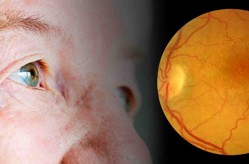
1. Fundus examination (ophthalmoscopy/fundus photo)
• The doctor with the help of a special device examines the macula.
• You can see:
o drusis - light yellowish deposits under the retina (the first sign of dry AMD),
o subtle changes in pigment epithelium.
2. Optical coherence tomography (OCT)
- The most accurate and sensitive method.
- Shows:
o changes in macular thickness,
o swelling,
o foci of degeneration,
o initial signs of a wet process.
- A non-toxic, painless study that allows you to detect AMD even before the onset of symptoms.
3. Amsler net (home test)
- A simple test for daily self-control.
- A person looks at a grid with straight lines - if they appear curved, blurred or disappear, it's a sign of macular involvement.
- However, this test does not detect AMD before the appearance of visual symptoms, only controls its progress.
4. Fluorescein angiography
- It is carried out with suspicion of a wet form.
- Detects newly formed blood vessels and fluid leaks that have not yet impaired vision.
When to be examined:
Category/Recommendations
Persons 50 + years: Every 1-2 years - mandatory retinal examination
In the presence of druze or heredity: Annually or more often
With complaints of image distortion, spots in the center of vision: Immediately - even with mild symptoms
SUMMARIZE:
To detect AMD before it affects vision, you need to undergo regular ophthalmological examination, especially after 50 years.
OCT is the most effective method of early diagnosis. Timely detection allows you to slow down the course of the disease and save vision for years to come.
To check whether there is age-related macular degeneration (AMD), you need to undergo a specialized examination by an ophthalmologist, which includes an analysis of the state of the macula - the central part of the retina responsible for acute central vision.
How to check if there is AMD:
1. Fundus examination (ophthalmoscopy)
- The main method of primary detection of AMD.
- Produced using a special microscope or fundus camera.
- The doctor can detect:
o drusy - yellowish deposits under the retina (sign of dry AMD),
o pigment epithelial changes,
o signs of newly formed vessels in wet form.
2. OCT (optical coherence tomography)
- The most accurate and sensitive method of diagnosing AMD.
- Creates a "slice" of the macula in real time.
- Allows you to see:
o fluid accumulation,
o thinning or thickening of the retinal layers,
o subnetwork formations,
o early degenerative changes even before symptoms appear.
3. Amsler mesh (vision distortion test)
- A simple test that can be performed even at home.
- The patient looks at the grid with straight lines (through one eye).
- If the lines seem wavy, curved or disappear - this is a sign of damage to the macula.
- Suitable for control, but not a substitute for examination.
4. Fluorescein angiography
- A dye (fluorescein) is injected into the vein, after which photos of the retina are taken.
- Allows you to see the pathological vessels and fluid leakage with wet AMD.
- Used when an active phase is suspected.
When should be examined for AMD:
Category Frequency of examinations
Persons aged 50 + years 1 once a year (even without symptoms)
People with AMD cases in the family 1 once a year or more
SUMMARIZE:
To check for AMD, you should consult an ophthalmologist for an examination of the fundus, OCT and additional tests.
AMD is best detected in the early stages - before vision loss, so regular examination after 50 years is extremely important.
Manifestations of age-related macular degeneration (AMD) depend on its form (dry or wet) and the stage of the disease. In the early stages, symptoms may be imperceptible, but with the progression of the disease, clear violations of central vision appear.
The main symptoms of AMD:
Initial manifestations (especially with dry form):
Slightly blurred or blurred vision
Difficult to read small print or work with details
There is a need for brighter lighting
Eye fatigue when reading
Typical manifestations (in later stages):
Curvature of straight lines (lines seem wavy - a key sign!)
Dark or gray spot in the center of vision
Difficulty recognizing faces
Blurred or darkened central vision
Deterioration of color perception
Contrast reduction (difficult to see at dusk)
Manifestations of wet (neovascular) AMD:
Sudden visual impairment (over days or weeks)
Dark or reddish spots (due to hemorrhages under the retina)
Distortion of objects and lines
Illusion of wavy images or curvature of geometry
What is stored at AMD:
Lateral (peripheral) vision - usually does not suffer.
A person can navigate in space, but does not clearly see what is right in front of him.
Symptoms requiring immediate medical attention:
There was a spot in the center of vision
Distortion of objects
Sudden visual impairment
Decreased visual acuity in one eye
SUMMARIZE:
Manifestations of AMD gradually affect central vision, making it difficult to read, write, recognize faces and drive.
The most noticeable sign is the curvature of straight lines and a spot in the center of vision. If even minor symptoms appear, you should immediately consult an ophthalmologist, because timely treatment can stop the loss of vision.
The most effective treatments for age-related macular degeneration (AMD) depend on its form:
Dry (atrophic) AMD - 85-90% of cases
Unfortunately, there is no specific treatment, but it is possible to slow the progression:
Recommended methods:
Balanced nutrition, rich in:
lutein and zeaxanthin (spinach, broccoli, eggs),
omega-3 fatty acids (fish),
vitamins C, E, zinc, copper.
Special antioxidant complexes (preparations based on the formula AREDS2):
slow the development of the disease by 25-30%.
Control of risk factors:
normalization of pressure, cholesterol,
complete cessation of smoking.
Regular observation by an ophthalmologist (1-2 times a year)
Wet (neovascular) form of AMD - ~ 10-15% of cases, but it is the most dangerous
The most effective methods of treatment:
Anti-VEGF injections (in the eye)
The most important and effective method:
Drug
Action
Aylia (Aflibercept)
Reduces swelling, stops the growth of new vessels
Wabismo (Faracamab)
Reduces swelling, stops the growth of new vessels
Usually introduced 1 once a month at the beginning, then - on an individual schedule.
Photodynamic therapy (PDT)
Combines the introduction of a photosensitive substance and laser activation.
It damages abnormal vessels, is rarely used - in cases where injections of anti-VEGF drugs are not effective.
Laser coagulation
With certain types of vascular lesions (non-central localization).
Less popular due to the risk of damage to healthy tissues.
LET'S UP:
Form of AMD
The most effective treatment
Dry
Antioxidants (AREDS2), nutrition, risk control
Moisture
Anti-VEGF injections (Aylia, Lucentis, Avastin) are the gold standard
Early detection is the key to effective treatment.
The sooner therapy is started, the more chances to maintain or improve vision.
It is impossible to completely cure age-related macular degeneration (AMD) today, but you can significantly slow down its development, and in the case of a wet form, stabilize or even improve vision if treatment is started on time.
Dry form of AMD:
Incurable, as it is associated with age-related depletion of macular cells.
But it can be slowed down:
intake of antioxidants (vitamins C, E, zinc, lutein, omega-3),
healthy diet and lifestyle,
blood pressure and cholesterol control,
regular observation by an ophthalmologist.
Purpose of treatment:
stop or slow down the progression of macular atrophy
maintain functional central vision for as long as possible
Wet form of AMD:
Not completely cured, but there are effective methods of control:
anti-VEGF injections (Aylia, Lucentis, Avastin)
some patients recover some vision
with regular treatment, vision can stabilize for years
Purpose of treatment:
stop the growth of pathological vessels
remove swelling
keep vision at the current level or slightly improve
So, is it possible to cure AMD?
Questions
Answer
Is it possible to completely get rid of AMD?
No, it is a chronic, incurable disease
Can vision be preserved?
Yes, with timely detection and treatment
Is it possible to improve vision in wet form?
Partly due to anti-VEGF injections
Is it possible to live fully with AMD?
Yes, if you follow the recommendations
SUMMARIZE:
AMD is not completely cured, but with early detection and proper treatment, you can effectively stop its progress and preserve vision.
The main thing is not to ignore preventive examinations after 50 years and consult an ophthalmologist with the slightest changes in vision.