

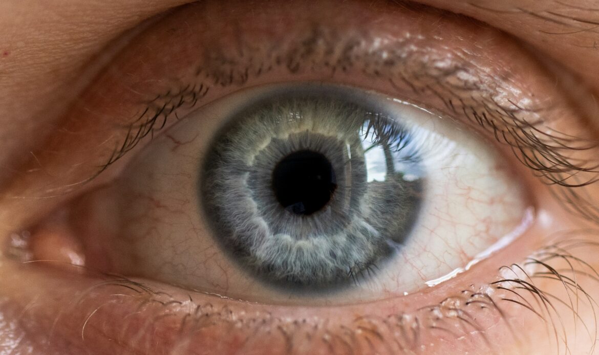
When a doctor examines the fundus, they can see the retinal blood vessels, but these are only part of the eye’s blood supply. Deeper lies the choroid, which nourishes the outer layers of the retina and the macula, responsible for sharp central vision. Because of its location, it cannot be directly examined, and disorders affecting it may remain asymptomatic for a long time. In this article, we will explain how this layer is structured and why its condition is important for preserving vision.
What Is the choroid of the eye?
The choroid, or choroidea, is the posterior part of the uveal tract (the middle layer of the eyeball). It is located between the sclera and the retina: large and medium-sized blood vessels lie in the outer layers, while the smallest capillaries are located directly beneath the retinal pigment epithelium. The choroid accounts for most of the eye’s blood flow.
Main functions of the choroid
- Nourishes the outer layers of the retina. It supplies photoreceptors (cells that respond to light) with oxygen and nutrients and removes metabolic waste products.
- Works together with the retinal pigment epithelium, which forms the outer blood-retinal barrier and allows only necessary substances to pass from the blood.
- Helps regulate eye temperature: blood carries away heat generated when light is absorbed.
- Supports clear vision. Melanin in the choroid absorbs excess light and reduces its scattering inside the eye.
Diseases of the choroid
Changes in the choroid may be associated with inflammation, impaired blood circulation, the growth of new blood vessels, or increased permeability of the vascular wall. The retinal pigment epithelium and retina are often affected as well.
Choroiditis
Choroiditis is inflammation of the choroid. It may be caused by infections (toxoplasmosis, tuberculosis), systemic inflammatory diseases (sarcoidosis, Behcet disease), and other conditions. The inflammation often also involves the retina (chorioretinitis).
Symptoms: decreased visual acuity, spots or “floaters” in the field of vision, blurred and distorted vision, and sometimes photophobia. Treatment depends on the cause: infectious cases may be treated with antibacterial, antiviral, or antiparasitic medications, while non-infectious cases may require anti-inflammatory or immunosuppressive therapy.
Choroidal neovascularization
This is the growth of abnormal new blood vessels that extend from the choroid beneath the retinal pigment epithelium or the retina itself. These vessels leak fluid or blood and disrupt retinal function. If the macula is affected, it becomes difficult to read, recognize faces, and distinguish fine details, while straight lines may appear distorted. This condition most commonly occurs in age-related macular degeneration and pathological myopia.
Treatment: intravitreal (into the vitreous body) injections of anti-VEGF medications, which block vascular growth factor. The treatment regimen and frequency of injections are determined by the doctor based on the examination results.
Central serous chorioretinopathy
In this condition, fluid accumulates beneath the retina, most often in the macular area. Increased permeability of the choroidal vessels and dysfunction of the retinal pigment epithelium play an important role. Straight lines may appear wavy, objects may seem smaller or larger, and the center of the image may appear blurred.
The condition often resolves on its own within a few months, but it requires monitoring. If the changes persist, recur, or threaten permanent vision loss, the doctor may recommend photodynamic therapy or another form of treatment. Corticosteroids (tablets, eye drops, ointments, or injections) can trigger or worsen this condition, so they should not be used without a doctor’s prescription. If you are already taking such medications, do not stop them on your own consult your doctor first.
Other vascular conditions of the eye
These conditions affect not the choroid, but the blood vessels of the retina and other parts of the eye:
- Central retinal artery occlusion. Vision usually disappears suddenly and painlessly in one eye. This is an emergency condition similar to a stroke: without oxygen, the retina is rapidly damaged, so immediate medical attention is required.
- Central retinal vein occlusion. Vision decreases less suddenly, but retinal hemorrhages and macular edema may occur.
- Diabetic and hypertensive retinopathy – damage to the retinal blood vessels caused by diabetes and hypertension.
- Vitreous hemorrhage (hemophthalmos) – bleeding into the vitreous body: sudden “floaters,” a veil or haze in front of the eye, and decreased vision.
- Subconjunctival hemorrhage – a bright red spot on the white of the eye. It may look alarming, but it is usually harmless and resolves within one to two weeks. However, if it occurs after an injury or is accompanied by pain or decreased vision, see a doctor.
How are vascular diseases of the eye diagnosed?
Depending on the symptoms, the doctor may recommend:
- ophthalmoscopy – examination of the fundus;
- optical coherence tomography (OCT) – a detailed cross-sectional view of the retinal layers;
- OCT angiography – visualization of blood flow in the vessels without the use of contrast dye;
- fluorescein angiography – after contrast dye is injected into a vein, it shows where blood vessels are leaking fluid;
- indocyanine green angiography – provides the best visualization of the choroidal blood vessels.
Not everyone needs all of these tests – the doctor selects them individually.
How are vascular eye conditions treated
Treatment depends on the diagnosis and begins with controlling the underlying condition: if vascular damage is associated with diabetes, hypertension, or atherosclerosis, it is important to keep blood sugar, blood pressure, and cholesterol levels within the normal range.
Then, depending on the indications, anti-inflammatory, antimicrobial, or immunosuppressive medications, anti-VEGF injections, photodynamic therapy, or laser treatment may be used, and in complex cases, surgery may be required. Sometimes regular monitoring is sufficient. Similar symptoms can occur with different diseases, so you should not diagnose yourself or take medications without a doctor’s prescription.
When should you see an ophthalmologist
Do not delay seeing a doctor if you notice decreased vision, distortion of straight lines, a dark spot in the center of your visual field, or unexplained blurred vision. If your vision worsens suddenly, especially in one eye, seek medical attention immediately. People with diabetes and hypertension need regular fundus examinations even if they have no symptoms; the frequency is determined by the doctor.
Eye health is not only about visual acuity today, but also about preserving your vision for the future. Schedule a consultation at Prostir Zoru Clinic – here, your eyes will be cared for as if they were our own.
Interesting facts about the blood vessels of the eye
From cancer drugs to preserving vision.
The idea that tumors grow by developing new blood vessels was proposed by Judah Folkman in the early 1970s. This led to the development of drugs targeting vascular endothelial growth factor (VEGF). One of them, bevacizumab, was initially used in oncology, and later ophthalmologists began using it in the eye as well. This transformed the treatment of wet AMD: previously, vision in such patients usually continued to deteriorate, whereas today it can often be stabilized and sometimes even improved.
A window into the blood vessels.
Research is currently being conducted into the use of AI for screening the risk of diabetes. AI can process large amounts of diagnostic data and assess retinal changes even in cases where a person has not yet noticed any deterioration in vision. The system analyzes a fundus image and, within a few minutes, determines whether an ophthalmologist’s consultation is needed, while complex cases can be reviewed by specialists even from another part of the world. The image can be taken by a family physician, endocrinologist, or another specialist, which is especially important in countries with limited access to healthcare. Researchers are also studying whether retinal images can be used to predict the risk of cardiovascular disease, stroke, and other conditions.

Kushnir Nataliya
Ophthalmologist, Candidate of Medical Sciences