
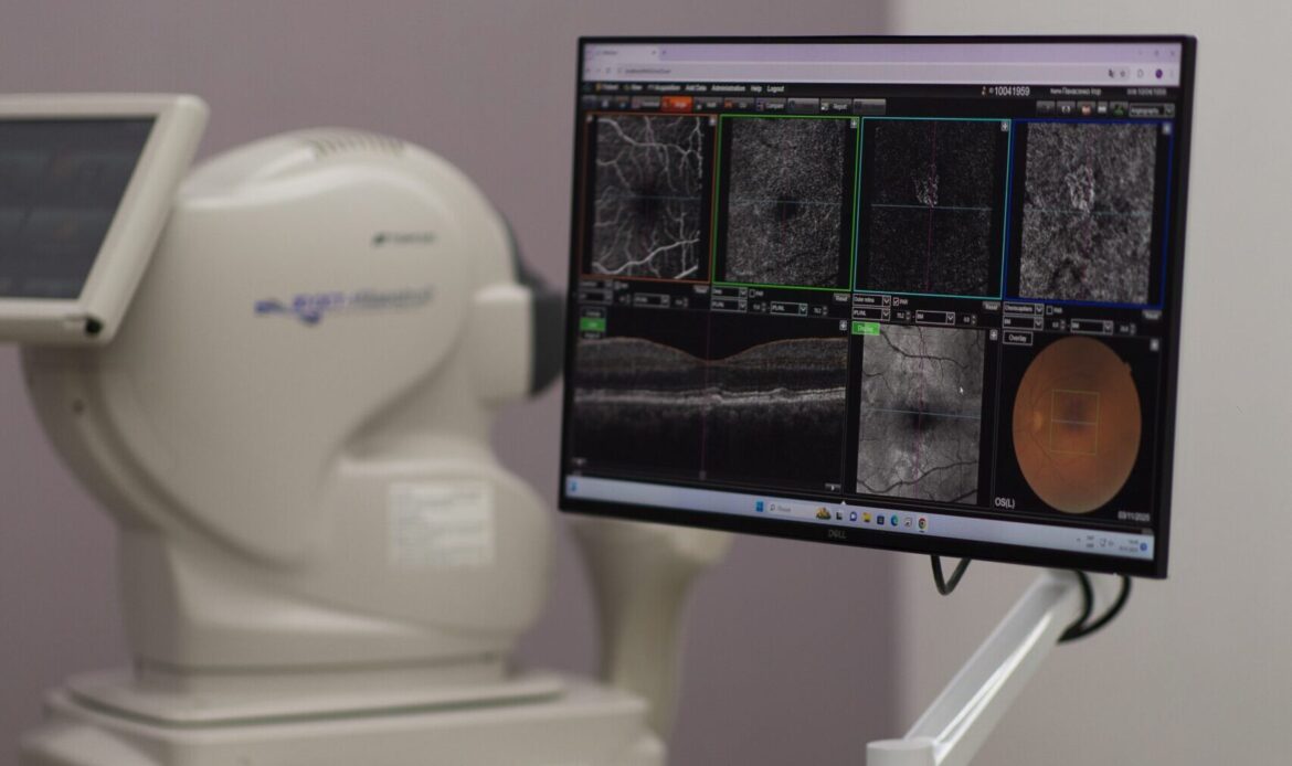
When a patient comes in for surgery, they want to be confident that their health is in safe hands. That is why we use high-tech surgical equipment that operates as a single integrated system — from the diagnostic office to the operating room. This seamless integration allows the surgeon to work with maximum precision while ensuring the patient feels safe and secure.
VERION Digital Marker M digital marker device (Alcon, USA, 2025)
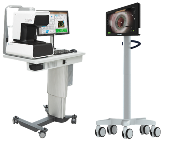
Preoperative calculation of the intraocular lens (IOL) is an extremely important stage in planning surgical intervention. To select the correct spherical power of the artificial lens tailored specifically to your eyes, measurements are performed using the ARGOS biometer. It serves as the initial component in the integrated equipment system provided by the leading American company Alcon.
VERION Digital Marker M is a digital navigation device that replaces traditional manual marking during intraocular lens (IOL) implantation surgery. Its primary function is to ensure precise positioning of the IOL.
Data from the biometer are transmitted to the VERION system via a network connection. These data are then synchronized with the eye image received from the surgical microscope. As a result, the surgeon sees virtual reference markers overlaid on the live image of the eye in real time. This allows for precise determination of the optimal positioning of the lens.
The system automatically adjusts alignment if the patient changes head position, further reducing the risk of inaccuracies. This approach significantly increases precision and minimizes the likelihood of errors that may occur with manual marking, especially in cases involving toric and multifocal IOLs, where exact orientation is critical.
In addition to navigation, the device allows reference images, surgical data, and videos to be stored on external media, which is useful both for medical documentation and for further analysis.
LuxOR Revalia ophthalmic surgical microscope (Alcon, USA, 2023)
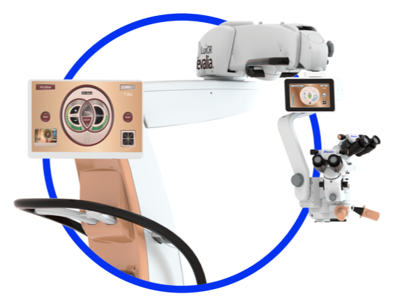
LuxOR Revalia is a modern ophthalmic surgical microscope that provides the surgeon with an exceptionally clear view of the eye during surgery. For the patient, this translates to greater safety, precision, and comfort, as the surgeon operates in fully controlled conditions.
Key features of this microscope:
Personalized lighting: The system adjusts the illumination so the surgeon can see all eye details without causing glare for the patient. This reduces discomfort and allows for more precise work.
Increased depth of focus: The surgeon does not need to constantly refocus the microscope, even if the eye’s position changes. This shortens the surgery time and reduces the risk of errors.
Stable red reflex (ILLUMIN-i® technology): Specialized lighting technology provides a clear view of the eye’s internal structures. For the patient, this means the surgeon can see better and operate with maximum accuracy.
Ergonomics and integration: The microscope easily integrates with other operating room systems, making the surgical process smoother and more controlled.
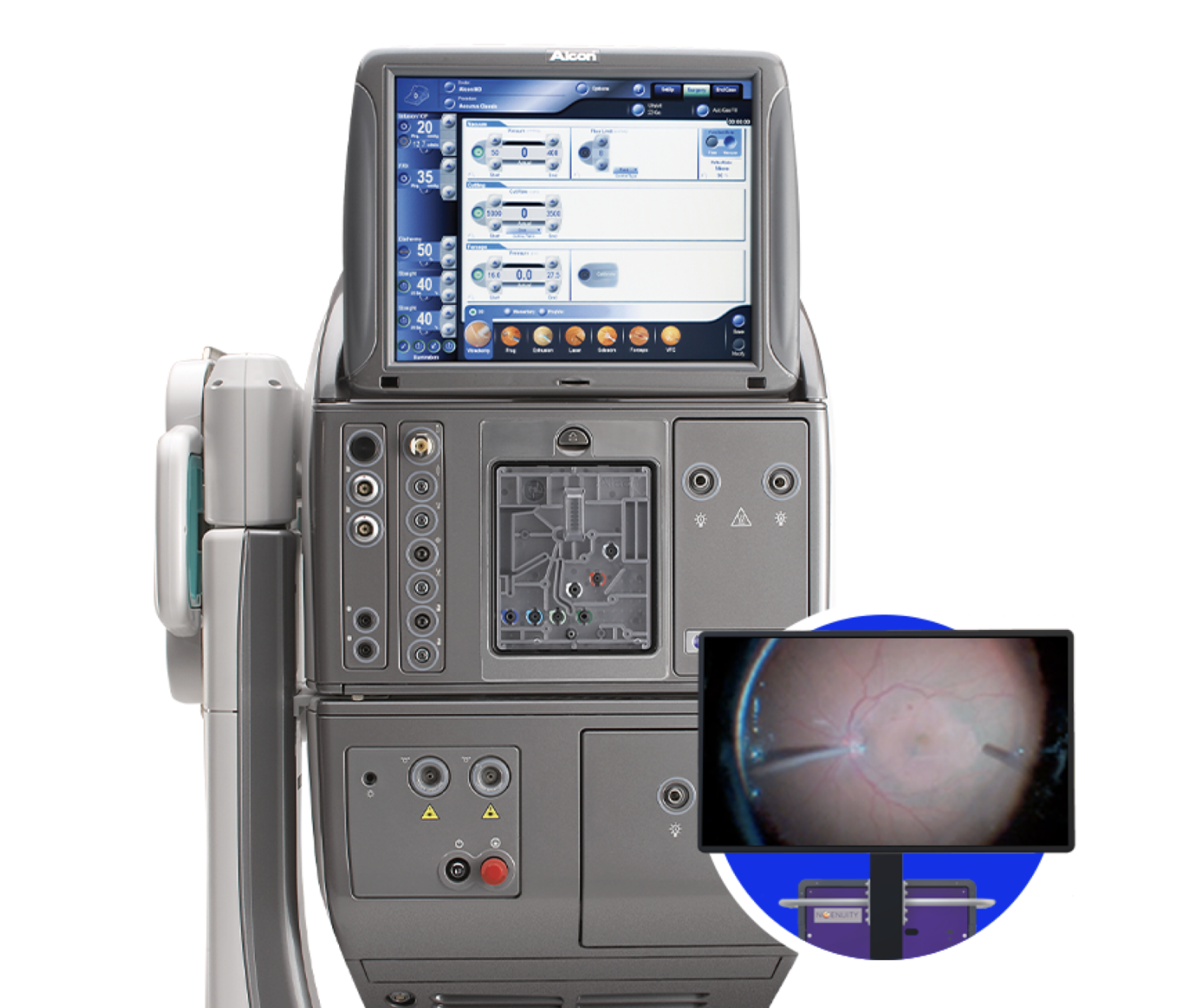
Constellation Vision System phacoemulsification and vitrectomy System (Alcon, USA, 2023)
Key features of the system:
High-speed ULTRAVIT® vitrectomy probes: Allow retinal and vitreous surgery with minimal tissue trauma.
Intraocular pressure (IOP) control: The system automatically maintains stable pressure inside the eye during surgery, which is critical for patient safety.
Cataract phacoemulsification: Advanced technology for removing a clouded lens followed by implantation of an artificial intraocular lens.
Integrated lighting (Xenon Illumination): Provides the surgeon with a clear view of the eye’s internal structures, reducing the risk of errors.
User-friendly control: A multifunction foot pedal and voice confirmation of settings allow the surgeon to operate quickly and without distractions.
What does this mean for you?
A modern level of safety and precision. Automatic control of your intraocular pressure provides the surgeon with stable operating conditions. Eye tissues recover faster thanks to minimal trauma. All of this allows for shorter surgery time, reduced risk of complications, and a predictable outcome, so you can feel confident in receiving high-quality and comfortable treatment.
NGENUITY 3D Visualization System (Alcon, USA, 2024)
NGENUITY is a digital 3D visualization system that enables ophthalmic surgeries to be performed in true three-dimensional format. For the patient, this ensures that the surgeon operates with maximum precision and reduced strain, while for the clinic, it provides opportunities for training and collaborative teamwork directly during the procedure.
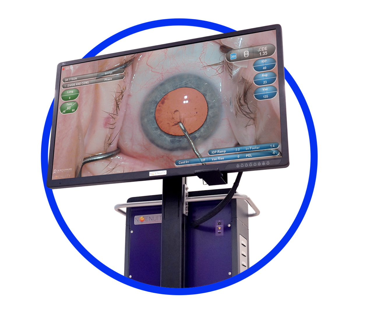
Key features of the system:
55-inch 3D 4K OLED display: Provides an exceptionally clear and detailed image.
3D stereocamera with high dynamic range: Captures all the nuances of the eye’s structure.
Fast image processing: The surgeon sees actions in real time without any delay.
Advanced technology surpassing analog microscopes: Up to 48% magnification, five times greater depth of field, and 42% higher resolution.
Photo and video recording: Allows high-resolution documentation of the surgery for analysis and training purposes.
What does this mean for you?
The surgery is performed in true 3D format, allowing the surgeon to see the eye in much greater detail than with a conventional microscope. Additionally, the system creates a comfortable working environment for the surgeon: they can operate with a straight back and raised head, reducing fatigue and improving focus. As a result, you benefit from a safer and higher-quality procedure.

Kushnir Nataliya
Ophthalmologist, Candidate of Medical Sciences